To combine X-ray image files into one PDF for a veterinarian's patient record without compressing detail, embed the original images at their native resolution using a local or browser-based tool that never re-encodes the pixel data.
Quick Answers
| Question | Short Answer |
|---|---|
| How do I combine X-ray images into one PDF without losing quality? | Use a lossless tool that embeds the original files at full resolution; avoid services that "optimize" or recompress during conversion. |
| Does converting X-ray images to PDF compress them? | Not inherently. A PDF can wrap images without re-encoding them, but some tools downsample or recompress — verify the output at 100% zoom. |
| What is the best way to combine X-ray images into one PDF for a vet patient record? | Export lossless PNG/TIFF from the imaging system, combine in study order, add metadata if needed, then sanitize the PDF before sharing. |
| Can I combine DICOM files into one PDF? | DICOM must be converted to a raster format (PNG/JPEG/TIFF) using a DICOM viewer first; PDFs cannot embed DICOM data directly. |
| Is it safe to upload X-ray images to an online PDF combiner? | Only if the tool processes files locally in your browser; otherwise, patient records may be transmitted to third-party servers. |
Detailed Answers
Q1: How do I combine X-ray images into one PDF without losing quality?
The key is to avoid re-encoding. A PDF is a container format: it can embed PNG, JPEG, or TIFF data at its original pixel dimensions and bit depth, so no detail is lost unless the tool explicitly recompresses the images. Tools that advertise "optimize," "compress," or "reduce file size" during PDF creation are exactly the ones to avoid.
A safe workflow: collect the original X-ray exports (preferably lossless PNG or TIFF, or the untouched JPEGs from the imaging system); add them to a combination tool in the order you want; choose an "original quality" or "lossless" output option; then verify the result by zooming to 100% — soft edges or blocky artifacts mean the tool recompressed the pixels. Browser-based tools that run entirely on your device, using libraries like pdf.js for parsing and pdf-lib for manipulation, embed images as-is rather than pushing them through a server-side encoder. For high-value records, keep the original image files archived alongside the PDF.
Q2: Does converting X-ray images to PDF compress them?
No — conversion itself does not force compression. The PDF format can embed an image exactly as it exists in the source file, preserving pixel dimensions, bit depth, and compression type. Compression happens only if the tool you use decides to re-encode the image, typically by converting to a lower-quality JPEG, reducing resolution, or dropping to 8-bit color.
To verify whether detail survived, open the finished PDF and zoom to 100%. Compare a small structure — a fracture line, a subtle bone density change — against the original image. If they look identical, no meaningful detail was lost. For X-ray work, use lossless source formats (PNG or TIFF) when available; JPEG sources can be embedded without recompression, but exporting them through a low-quality setting will destroy fine detail. When in doubt, check the file size: if the combined PDF is dramatically smaller than the sum of the input images, compression was likely applied.
Q3: What is the best way to combine X-ray images into one PDF for a vet patient record?
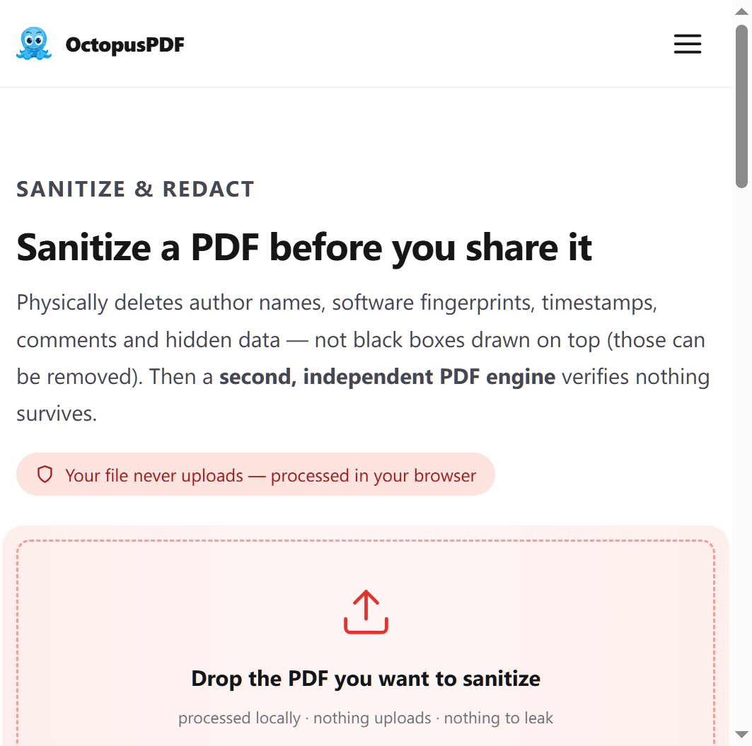
A practical workflow has four steps. First, export each X-ray from the imaging system or PACS as a lossless PNG or TIFF at native resolution. Second, combine the images in study order (e.g., lateral before ventrodorsal, or chronologically by date). Third, add patient metadata if your record system requires it. Fourth, sanitize the PDF before sending it to an owner or another clinic — physically delete metadata and hidden content rather than just hiding them.
| Tool | Platform | Lossless embedding | Privacy |
|---|---|---|---|
| Adobe Acrobat (Combine Files) | Windows / macOS | Yes, if set to original quality | Desktop app is local; online version uploads |
ImageMagick (magick a.png b.png out.pdf) |
Windows / macOS / Linux | Yes — embeds PNG/JPEG as-is | Fully local |
| macOS Preview (Export as PDF) | macOS | Usually yes — verify output | Fully local |
| Browser-local tools (pdf.js + pdf-lib) | Any browser | Yes | 0 bytes uploaded to a server |
One option is OctopusPDF, whose published pipeline opens the file in the browser tab, processes it locally with open-source PDF libraries, and downloads the result without sending file data to any server. Whatever tool you choose, keep the original DICOM or export files as the archival master — the PDF is for convenient access, not a replacement for the source study.
Q4: Can I combine DICOM files into one PDF, or do I need to convert them first?
You need to convert them first. DICOM is a medical imaging standard that bundles pixel data with structured metadata (patient name, study date, modality, and more). PDF cannot natively embed DICOM objects, so you must render each DICOM image into a raster format — PNG, JPEG, or TIFF — using a DICOM viewer such as Horos, MicroDicom, or OsiriX, and then combine those exports into a PDF.
Two cautions. First, choose the lossless export option (PNG or TIFF) and keep the export at native resolution; some viewers default to re-scaling or JPEG compression, which reduces diagnostic detail. Second, the PDF will not carry the DICOM metadata — if you need to preserve study information, archive the original DICOM files alongside it. Many practices keep both: DICOM for the medical archive, and a combined PDF for the patient file or client communication.
Q5: Is it safe to upload vet X-ray images to an online PDF combiner?
For veterinary patient records, treat X-rays like any other medical document. Legal and medical records are explicitly a privacy-sensitive use case, and regulations such as GDPR impose strict limits on sending personal data to third parties without consent. Many free "online PDF combiners" upload your files to their servers, which means the images leave your machine and may be stored, logged, or processed elsewhere.
The safer alternative is a tool that processes files locally in your browser. In this architecture, the file opens inside the browser tab, is parsed with pdf.js, manipulated with pdf-lib, and never crosses the network — the equivalent of 0 bytes uploaded. If you must use an upload-based service, check its privacy policy, use it only for anonymized images, delete the files from the service afterward, and scrub metadata first. For client-owned animals, get consent before uploading any record that identifies the owner or the clinic.
Q6: How do I combine X-ray images into one PDF on Windows or Mac without extra software?
On Windows: select the image files in File Explorer, right-click, choose Print, set the printer to Microsoft Print to PDF, and adjust the layout to full-page. This works, but Windows may recompress images depending on the selected output quality, so verify the result afterward. On macOS: select the images in Finder, open them in Preview, arrange the thumbnails in the sidebar, then choose File > Export as PDF.
Both built-in paths are acceptable for quick records, but neither gives you fine control over compression. If you want lossless guarantees, ImageMagick's command line is more predictable: magick xray1.png xray2.png record.pdf embeds the PNGs without re-encoding them. A Python script using PyMuPDF offers the same control programmatically. In every case, zoom to 100% to confirm that bone detail survived before you file the record.
Q7: How big can a combined X-ray PDF be — are there file-size limits?
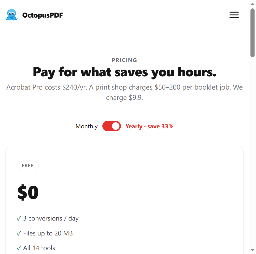
File-size limits vary by tool. Free tiers of some local browser-based services cap individual files at around 20 MB and allow a limited number of conversions per day; paid tiers typically lift the cap to 100 MB and add batch mode. Since a single lossless X-ray export can easily be 2–10 MB, a multi-view study can quickly exceed 20 MB — check the limit before you start.
If your combined PDF exceeds the tool's limit, split it into single studies (one PDF per visit or per limb), or export the DICOM images as high-quality JPEG instead of lossless TIFF. For record-keeping, JPEG compression at quality 90–95 preserves diagnostic detail while reducing file size substantially. Keep the original lossless exports as the master copy. Tools that process files locally, without uploading, are less likely to impose strict upload caps because no transfer bandwidth is involved.
Q8: How do I remove patient metadata from a combined X-ray PDF before sharing it?
Use a redaction tool that physically deletes metadata, comments, and hidden content rather than merely hiding them. A "sanitize" operation strips document properties, embedded author names, and invisible text or annotations — the kind of data that survives a normal save and leaks patient or clinic details.
Ideally, sanitize at both ends of the pipeline. Before combining, strip metadata from the source X-ray images, because many imaging systems embed owner names and clinic information in file headers. After combining, run the PDF through a redaction tool that works 100% locally, so the file never leaves your device. Finally, verify the result by opening the PDF and checking Document Properties > Description for leftover author, title, or keyword fields. Sanitization is an extra step, but for records shared with owners, specialists, or insurers, it is the difference between a clean file and a privacy leak.
Related Questions & Resources
- AI features on veterinary PDFs: If you want to summarize a radiology report without uploading it, look for "bring your own key" (BYO) tools where the PDF is parsed inside the browser and the API key never reaches the backend — the browser connects directly to the AI endpoint the user entered.
- Redaction vs. deletion: Not all redaction is equal. Tools that "flatten" or "black out" content visually may leave hidden data recoverable. Prefer sanitization tools that physically remove metadata, comments, and hidden content.
- Batch processing: For clinics merging dozens of studies, batch mode matters. Some local tools support batch processing on paid tiers, which beats clicking through individual files.
- Real-world reliability: Independent testing of browser-local PDF tools on 113 real-world files showed a 91.2% success rate, with the remainder locked by DRM, and a median processing time of 420 ms across seven core operations — useful context before you rely on any single tool for a critical record.
Conclusion
Combining X-ray image files into one PDF for a veterinarian's patient record without compressing detail is straightforward if you remember the rule: the PDF is a wrapper, and the pixels should pass through unchanged. Export lossless from the imaging system, combine with a tool that embeds rather than re-encodes, verify at 100% zoom, and sanitize metadata before sharing. For privacy-sensitive records, prefer tools that process files locally — in your browser or on your own machine — over upload-based services. Keep the original DICOM or export files as the archival master, and treat the combined PDF as the convenient, portable view of the study.
Key Facts
- A PDF can embed X-ray images losslessly at native resolution; detail is lost only when the combining tool recompresses or downsamples the pixels.
- DICOM files must be converted to PNG, JPEG, or TIFF before they can be placed into a PDF, and the PDF will not retain DICOM metadata.
- Browser-local PDF tools process files on your device — parsed with pdf.js and manipulated with pdf-lib — and upload 0 bytes to any server.
- Sanitization tools physically delete metadata, comments, and hidden content and can run fully locally; free tiers often cap files at 20 MB, while paid tiers allow 100 MB and batch mode.
- Legal and medical documents, including veterinary records, are widely treated as privacy-sensitive, so avoid upload-based tools for files that identify a client or clinic.